HydroPhotonics®

The Optimal Wavelength

  • Waterlase’s proprietary 2780 nm Er,Cr:YSGG wavelength provides optimal energy absorption into the water and Hydroxyapatite found in enamel, dentin, and bone.
  • Laser photons energize atomized water spray and micro-droplets native to interstitial spaces in tissue, creating rapid expansion/micro-explosions in the water.
  • This photomechanical ablation is termed HydroPhotonics.
    With no pain in most cases*, the result is rapid, precise, and clean removal of target tissues — without heat damage, fractures or smear layer created by dental burs.
  • Waterlase laser energy has been shown to have an anti-bacterial effect in root canals, reducing E. faecalis by 99.71%.**
Laser Wavelength Absorption Chart

ER,CR:YSGG

Optimal Absorption

Waterlase’s proprietary Er,Cr:YSGG 2780 nm wavelength has optimal absorption in both water and Hydroxyapatite for cool and efficient cutting in both hard and soft tissue. This is the therapeutic “Goldilocks” zone.

  • SOFT TISSUE: Er:YAG water absorption is too high. Er,Cr:YSGG penetrates water containing tissues 300% deeper than Er:YAG, which results in efficient cutting with better hemostasis, deeper coagulation and less bleeding.***
  • HARD TISSUE: Hydroxyapatite absorption of CO2 wavelengths are too high, resulting in thermal ablation, melting, heating and carbonization depending on technique, tissue, and settings.†

Histological Comparison

Histologic Comparison

Porcine gingival tissue histology from Figure 4. of Kawamura, Rie et al. “Ex Vivo Evaluation of Gingival Ablation with Various Laser Systems and Electroscalpel.” Photobiomodulation, photomedicine, and laser surgery vol. 38,6 (2020): 364-373. doi:10.1089/photob.2019.4713

* Based on clinician-reported outcomes. ** Gordon, W. et al. “The Antimicrobial Efficacy of the Er,Cr:YSGG Laser with Radial Emitting Tips on Root Canal Dentin Walls Infected with e. Faecalis.” JADA 138, July (2007): 992–1002. *** 
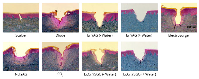
1. Cercadillo-Ibarguren, I., et al. “Histologic Evaluation of Thermal Damage Produced on Soft Tissues by CO₂, Er,Cr:YSGG and Diode Lasers.” Medicina Oral Patología Oral y Cirugia Bucal 15, no. 6 (2010): e912–18. 2. Romeo, U., et al. “Histological in Vitro Evaluation of the Effects of Er:YAG Laser on Oral Soft Tissues.” Lasers in Medical Science 27, no. 4 (2012): 749–53. † Christensen, Gordon J. “First Look: Solea CO2 Hard and Soft Tissue Laser.” CLINICIANS REPORT, Volume 8 Issue 4, April 2015, Pages 1, 3 & 4 1. Hossain, M. et al. “Analysis of Surface Roughness of Enamel and Dentin after Er,Cr:YSGG Laser Irradiation.” Journal of Clinical Laser Medicine & Surgery 19.6 (2001): 297–303. 2. Baygin, O. et al. “The Effect of Different Enamel Surface Treatments on the Microleakage of Fissure Sealants.” Lasers in Medical Science 27.1 (2012): 153–60. 3. Kilinc, E. et al. “Thermal Safety of Er:YAG and Er,Cr:YSGG Lasers in Hard Tissue Removal.” Photomedicine and Laser Surgery 27.4 (2009): 565–70. 4. Fried, D. et al. “Mechanism of Water Augmentation during IR Laser Ablation of Dental Enamel.” Lasers in Surgery and Medicine 31, no. 3 (2002): 186–93. 5. Vogel, A. and V. Venugopalan. “Mechanisms of Pulsed Laser Ablation of Biological Tissues.” Chemical Reviews 103, no. 2 (2003): 577–644. 6. Majaron, B. “Thermo-Mechanical Laser Ablation of Hard Dental Tissues: An Overview of Effects, Regimes, and Models.” SPIE 3593, January (1999): 184–95. 7. Türkün, M. et al. “Bactericidal Effect of Er,Cr:YSGG Laser on Streptococcus Mutans.” Dental Materials Journal 25.1 (2006): 81–6.
Made in the USA / YSGG Inside

Contact us today

Learn what Waterlase can do for your practice. 

Waterlase iPlus™

WATERLASE

iPLUS